Bone marrow stromal cells (MSC) are pluripotent progenitor cells that have the capability to migrate towards lesions and induce or facilitate site-dependent differentiation in response to environmental signals. Using in vivo MR imaging, we studied MSC transplantated into adult rats with a cortical photochemical lesion.
|
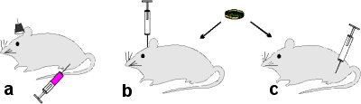
The induction of cortical photochemical lesion with rose bengal and light beam interaction (a), The intracerebral administration of MSC (b), The i.v. administration of teh MSC (c). |
MSC were isolated from rat bone marrow by adherence to plastic. After in vitro expansion, the cells were co-labeled with superparamagnetic iron-oxide nanoparticles (Endorem, Guerbert Laboratories, France) and BrdU (5 mM) 48 hours prior to transplantation and administered either intracerebrally into the contralateral hemisphere (0.3 million cells in 3ml PBS; n=12) or i.v. into the femoral vein (2 million cells in 0.5 ml PBS; n=8). A photochemical lesion was induced by rose bengal/light beam interaction 24 hours prior to transplantation. MR images were taken weekly using a 4.7 T Bruker spectrometer. Rats were sacrificed 4 weeks following transplantation, and the fate of transplanted cells in the CNS was analyzed immunohistochemically. The cells preferentially migrated into the lesion, and subsequently some of the cells that entered the lesion expressed the neuronal marker NeuN. In animals without a lesion, the majority of intracerebrally injected cells remained in the close vicinity of the needle track.
|
|
|
|
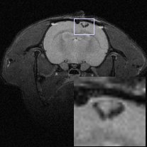
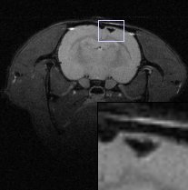
| T2-weighted images of cortical photochemical lesion 12 hours (left), 7 days (middle) and 30 days (right) after the intravenous application of MSC labeled with nanoparticles. | ||
Starting 7 days after transplantation and persisting for 4 weeks, MR images showed a hypointense signal in the lesion, indicating the migration of cells to the lesion. Hypointensity was also found in the lesion after i.v. injection of labeled cells. Prussian blue staining confirmed the presence of iron-oxide-labeled cells in the lesion site. The study demonstrates that iron-oxide nanoparticles can be used to track implanted stem cells in the CNS.
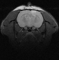
| The MSC labeled with nanoparticles and BrdU implanted directly into the cortical photochemical lesion. | 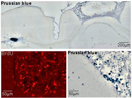 |