Presently, a great deal of attention is being paid to the research and clinical applications of MRI and MRS in biomedicine. In collaboration with the MR Unit of the Department of Diagnostic and Interventional Radiology at the Institute for Clinical and Experimental Medicine, we are investigating diffusion and perfusion in the brain and studying tissue metabolite concentrations by use of in vivo MR spectroscopy, utilizing an experimental spectrometer Bruker 4.7 Tesla.
|
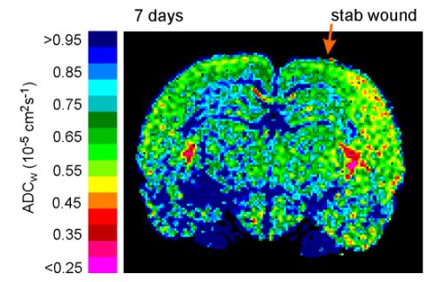
Pseudocolor image showing the ADCW map of a rat brain 7 days after a cortical stab wound. Note that ADCW is lower in the entire cortex of the wounded hemisphere than in the contralateral hemisphere. |
Part of our research involves the investigation of brain function in both humans and suitable animal models. The animal models focus on ischemic lesions, hypoxia, brain injury, hydrocephalus and transgenic animals. The MR data are compared with results obtained from diffusion analyses performed by the real-time iontophoretic method using ion-selective microelectrodes. The ultimate aim of this project is to make possible greater utilization and finer processing of MR spectra and MR images and to find a link between data obtained using MR imaging and changes in extracellular space diffusion parameters revealed by iontophoretic measurements.
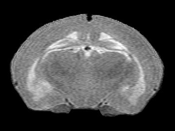 |
Normal brain (left) and Inherited hydrocephalus (right) in a 19-day-old rat: T2 - weighted images. | 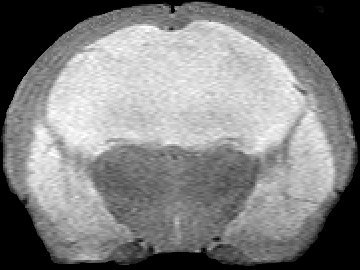 |