Extracellular space volume and geometry - factors
affecting diffusion in the CNS in health and disease
Diffusion in the ECS obeys Fick’s law, subject to three
important modifications. First, diffusion in the ECS is constrained by the
restricted volume of the tissue available for diffusing particles, i.e. by the
extracellular volume fraction (a). Second, the free
diffusion coefficient, D, is reduced by the square of the tortuosity (l)
to an apparent diffusion coefficient ADC = D/l2,
due to an increase in the path length for diffusion between two points and
because the diffusing substance encounters membrane obstructions, glycoproteins,
macromolecules of the extracellular matrix, charged molecules and glial cell
processes. Third, the diffusion of substances may be affected by nonspecific
uptake, k´, a factor describing the loss of a substance across cell membranes.
If we incorporate factors a, l
and k´ into Fick’s laws, diffusion in the CNS is described fairly
satisfactorily.
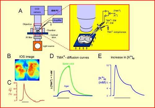
Set-up for simultaneous measurements of light transmittance (IOS), ECS
diffusion parameters and/or [K+]e changes. B: Example of
an IOS image. C: Changes in light transmittance. D: TMA+
diffusion curves recorded in spinal cord and agar. E: Changes in [K+]e
evoked by neuronal activity.
Studies in our laboratories have shown that the extracellular volume fraction
changes during development, being about twice as large in the cortex and corpus
callosum of newborn rats as in adults. The large ECS in the developing CNS might
allow for the more effective diffusion of macromolecules, such as growth factors
and cytokines. The reduction in ECS volume fraction with increasing age
correlates well with gliogenesis and myelination. Changes in the membrane
currents of glial cells associated with myelination have also been correlated
with ECS diffusion parameters.
Extracellular space diffusion parameters were studied during aging. Aged rats
were classified according to their performance during place learning, and two
groups, good and bad learners, were selected. Diffusion measurements were
performed along three orthogonal axes. The volume fraction and nonspecific
uptake were significantly lower in both aged groups than in young adults. In
young adults and good learners, anisotropy was found in the hippocampus; the
anisotropy was lost in bad learners. The loss of anisotropy in the hippocampus
of aged bad learners corresponded to the disorganization of glial processes and
a loss of extracellular matrix (fibronectin and chondroitin sulfate proteoglycan).
The significant differences in diffusion parameters between good and bad
learners in the CA3 and DG regions of the hippocampus may affect LTP, memory and
learning.
 |
 |
|
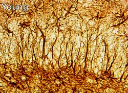
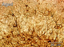
GFAP staining in the hippocampus of a young adult
and aged rat showing the disorganization of parallel glial processes
during aging. |
During hypoxia, our experiments have revealed that a
dramatically decreases, while l significantly
increases. The time course of the changes is about ten times slower in neonatal
rats than in adults, correlating with the well-known resistance of the immature
CNS to anoxia. These changes in diffusion parameters during and after ischemia
enhance the accumulation of substances, contributing to brain damage and
hindering the influx of metabolic substances during any subsequent reperfusion.
Diffusion-weighted MRI studies have shown that the apparent diffusion
coefficient of water is dramatically reduced during anoxia and ischemia, with a
time course that correlates well with the observed changes in a
and l. Further experiments are aimed at elucidating
the mechanism underlying these changes in water diffusion during anoxia.
|
|
|
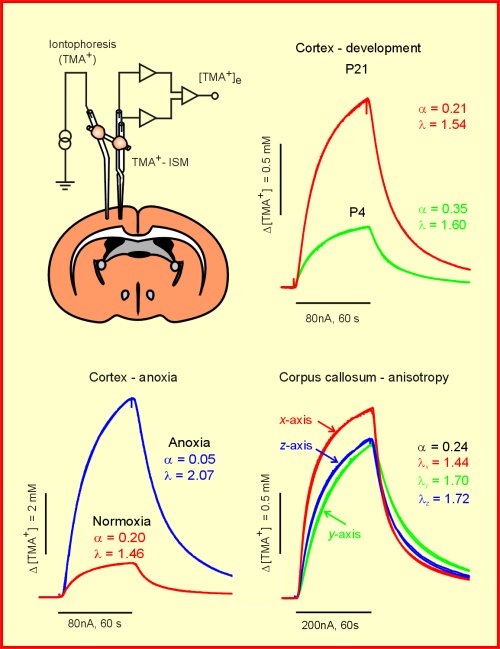
Scheme of the experimental arrangement for measuring
ECS diffusion parameters using the real-time iontophoretic method and TMA+-selective
microelectrodes and typical recordings obtained during development at
postnatal days 4 and 21, during normoxia and anoxia, and in the corpus
callosum under anisotropic conditions. |
Brain injury, with consequent neuronal death and astrogliosis, results
in changes in CNS architecture. Changes in diffusion parameters in experimental
models of injury and regeneration such as stab wounds and radiation necrosis
have been compared with histopathological changes in order to elucidate their
possible mechanisms. Experimental animal models have revealed that in the
vicinity of the injury, ECS volume and the apparent diffusion coefficients of
both water (ADCw) and tetramethylammonium (ADCTMA) are
decreased due to cell death and astrogliosis. In the lateral region of the
ipsilateral cortex, where no changes in ECS volume are found, prominent
increases in extracellular matrix expression (chondroitin sulfate proteoglycan)
are seen along with decreases of both ADCw and ADCTMA.
This shows that the apparent diffusion coefficient of water is affected by
diffusion barriers resulting from an increase in extracellular matrix.
Following intracerebral bacterial inoculation, acute inflammation and
increased blood-brain barrier permeability occurs, resulting in moderate changes
in ECS diffusion parameters. More dramatic changes, particularly an increase in
extracellular space volume, were seen in our studies utilizing an animal model
of the demyelinating disease multiple sclerosis, experimental autoimmune
encephalomyelitis, induced by an injection of myelin basic protein.
Recent experiments in the Department have used the 6-OHDA-lesion rat model of
Parkinson’s disease to study two different grafting techniques and
their influence on ECS diffusion parameters. Micrografting involves the
transplantation of fetal dopaminergic cells into a number of small deposits in
the striatum, while macrografting uses a single, larger deposit. We found a
functional recovery, good survival of tyrosine hydroxylase-positive cells and
astrogliosis in rats 3-5 months after grafting. Grafts were localized by T2 or
diffusion-weighted NMR, and ECS diffusion parameters were investigated in the
striatum. Tortuosity increased in the grafts and the adjacent tissue. The
increase in ECS diffusion barriers corresponded to the astrogliosis in and
around the grafts. The increased extracellular tortuosity therefore suggests an
impediment to dopamine diffusion from the grafts into the lesioned striatum.